From a rodent’s optic nerve and the venomous fangs of a small tarantula to auto-fluorescing hairs on a leaf, the images winning the Nikon Small World Photomicrography Competition are luminously colorful works of art revealing the artistic side of science.
For its 49th edition of photos showing the beauty of a world that can only be seen with a microscope, each image was judged not only for its scientific technique but also its artistry. The winners were selected from among 1,900 entries from 72 countries and a wide array of scientific disciplines.
Rodent optic nerve head showing astrocytes (yellow), contractile proteins (red) and retinal … [+]
Photo: Hassanain Qambari & Jayden Dickson – Nikon Small World 2023
This year’s first place was awarded to Hassanain Qambari, assisted by Jayden Dickson of the Lions Eye Institute (a center for ophthalmological care and scientific research), for his vivid image of a rodent optic nerve head showing astrocytes (yellow), contractile proteins (red) and retinal vasculature (green). The colorful image provides an important contribution to the study and reversal of diabetic retinopathy, which affects one in five persons with diabetes worldwide.
“The competition not only celebrates the participants’ hard work and passion but may also draw and inspire young scientists to pursue a career in STEM. It certainly inspired me,” said Qambari.
All the images of the top 10 winners, honorable mentions and Images of Distinction can be found on the Nikon Small World website.
5th Place, Norfolk, United Kingdom
Photo: David Maitland – Nikon Small World
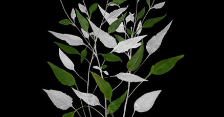
Auto-fluorescing defensive hairs covering the leaf surface of Eleagnus angustifolia exposed to UV light.
4th Place, Venomous fangs of a small tarantula, Medienbunker Produktion Bendorf, Rheinland Pfalz, … [+]
Photo: John-Oliver Dum – Nikon Small World 2023
The venomous fangs of a small tarantula inspire terror at this size.
Image of Distinction, Bee, TCDD Teknik Müh. Müş. A.Ş. Ankara, Çankaya, Turkey
Photo: Yusuf Ziya Öztürk – Nikon Small World 2023
Honorable Mention, Trichenella worm, Bellevue, Washington, USA
Photo: Dr. Nathan P. Myhrvold – Nikon Small WOrld 2023
This is a trichinella cyst in pork muscle. (Trichinella is a parasitic worm known to cause trichinosis).
Image of Distinction, Hibiscus flower bud, Bangalore, Karnataka, India
Photo: Raghuram Annadana – Nikon Small World
Stamen and stigma develop inside a Hibiscus flower bud in Bangalore, India.
Image of Distinction, Crab spider (Thomisus onustus), Saint Lys, Haute-Garonne, France
Photo: Sébastien Malo – Nikon Small Worl 2023
6th Place, Slime mold, Vacaville, California, USA
Photo: Timothy Boomer – Nikon Small World 2023
Slime mold (Comatricha nigra) shows its capillitial fibers through its translucent peridium.
12th Place, Cuckoo wasp, Tanta University, Egypt
Photo: Sherif Abdallah Ahmed – Nikon Small World
Cuckoo wasp stands on a flower at Tanta University Department of Zoology in Tanta, Egypt.
Image of Distinction, Ostracods and algae (Cladophora), Nassau Community College Department of … [+]
Photo: Frank Reiser – Nikon Small World 2023
Ostracods flit through algae, surrounded by bubbles.
7th Place, Mouse embryo, University of Geneva Department of Genetics and Evolution Geneva, … [+]
Photo: Dr. Grigorii Timin & Dr. Michel Milinkovitch, Nikon Small World
8th Place, Caffeine crystals, University of Georgia Athens, Georgia, USA
Photo: Stefan Eberhard
Image Of Distinction, Tapeworm, National Autonomous University of Mexico
Photo: Arturo Calderón, Dr. Miguel Tapia-Rodríguez & Dr. Juan Pedro Laclette – Nikon Small World
Muscle architecture of an evaginating tapeworm (Taenia crassiceps cysticercus) at National Autonomous University of Mexico (UNAM) Department of Immunology, Mexico City.
Image of Distinction, Cabbage butterfly eggs, Medienbunker Produktion Bendorf, Rheinland Pfalz, … [+]
Photo: John-Oliver Dum – Nikon Small World 2023
Image of Distinction, Marine organism (Pyrocystis lunula, Dinophyceae), University of Applied … [+]
Photo: Frank Fox – Nikon Small World
Image of Distinction, Mushroom gills (sporangiophores) showing sporophores, Issaquah, Washington, … [+]
Photo: Charles Krebs – Nikon Small World 2023
Image of Distinction, Leaf epidermis stomata (Stromanthe sp.), Suwalki, Podlaskie, Poland
Photo: Marek Miś – Nikon Small World 2023
Image of Distinction, Fly caught in a Venus flytrap, LaVision, Miltenyi Biotec Company Department of … [+]
Photo: Dr. Simon Frederik Merz Dr. Lea Bornemann, Dr. Ewa Patrycja Smajek – Nikon Small World
Image of Distinction, Buckthorn trichomes, Rotterdam, Zuid-Holland, Netherlands
Photo: Walter Machielsen – Nikon Small World 2023